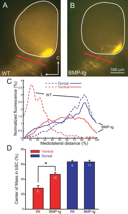
Figure 2.
Ventral retinal axons in BMP transgenics are misplaced in the brachium of the Superior Colliculus (bSC) at P8. A. Axons originating in ventral retina of WT mice enter the SC from the medial edge of the bSC in WT mice. B. In BMP-transgenic mice, ventral axons lose their confinement and spread laterally in the bSC. C. The difference in the distribution of RGC axons from ventral (red) retina in the bSC is shown as normalized fluorescence across the width of the bSC (red bar in Fig. 2A and 2B) for representative examples of WT (dashed line) and BMP-transgenic mice (solid line). The axon distributions in the bSC resulting from dorsal (blue) injections are similar in the two genotypes. D. These results are summarized as the mean position of the center of mass of the fluorescent distribution of labeled RGC axons from ventral (red) or dorsal (blue) injections across the width of the bSC, with the number of animals indicated above each bar. The mean centers of mass for the ventral retinal injections retina are significantly different between the two genotypes (*; p<0.001).

