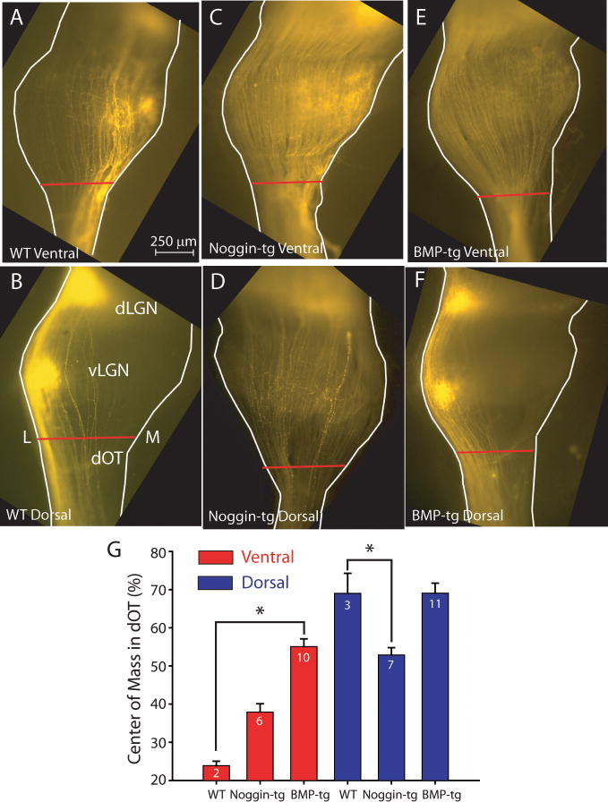
Figure 4.
Axons in the ascending ‘delta’ of the Optic tract (dOT) are missorted at P8 in BMP and Noggin transgenics. A, B. Axons originating from ventral or dorsal retina in WT mice travel along the medial (M), or lateral (L) edges of the optic tract. C, D. In the Noggin-transgenic mice, axons from both ventral and dorsal retina lose their confinement in the dOT, though the disturbance of the dorsal axons is more severe E, F. In BMP transgenic mice, the ventrally originating axons lose their restriction to medial dOT, while those from dorsal retina remain on the lateral side. G. Summary quantification of the position of the center of mass of fluorescent label across the medial-lateral width of the dOT (red bar in Fig. 4B) in WT, BMP transgenic and Noggin transgenic mice for dorsal (blue) and ventral (red) retinal injections. In BMP-transgenic mice, only injections from ventral retinal show an inappropriate distribution in the dOT, where as in Noggin transgenics, dorsal RGC axons are the most inappropriately distributed (*; p < 0.01).

