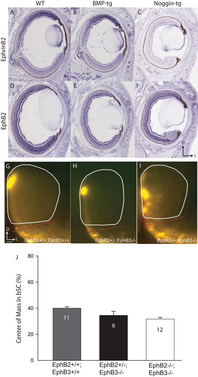
Figure 9.
In situ hybridization reveals altered expression patterns of EphB2 and ephrinB2 in the retina of BMP-tg and Noggin-tg mice, but ventral axons are not missorted in EphB2/B3 mutant mice. A. In WT mice, ephrinB2 is expressed in a high-dorsal to low-ventral gradient. B. The ephrinB2 expression pattern is not altered in BMP-tg mice. C. EphrinB2 expression is dramatically suppressed in Noggin-tg mice. D. In WT mice, EphB2 shows a high-ventral to low-dorsal expression pattern. E. EphB2 expression is suppressed in BMP-tg, and no obvious gradient is present. F. EphB2 expression lacks a high-ventral to low-dorsal pattern in Noggin-tg mice, though average expression levels are normal. G-I. At P8, relative to controls (G), ventral RGC axons in EphB2+/-;B3-/- (H) and EphB2-/-;B3-/- mice (I) are not missorted in the brachium of the SC (red bar in G and H). Summary quantification of center of mass for ventral injections in the bSC of WT and EphB2/B3 mutants. The sorting of EphB2/B3 double mutants is slightly better (with a smaller Center of Mass) than controls. D, dorsal; L, lateral; C, cadual; Scale bar: 100μm in G-I.

