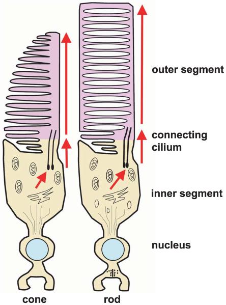
Figure 5.
Morphology of vertebrate cone and rod photoreceptors. Each includes an inner segment and an outer segment connected by a cilium. The inner segment contains the cell's biosynthetic machinery. Phototransduction takes place in the outer segment. In the rods, the outer segment contains free-floating membrane-bound disks. In cones, the disks remain continuous with the plasma membrane. Modified from Perkins BD, Fadool JM (2010) “Photoreceptor structure and development analyses using GFP transgenes.” Methods in Cell Biology 100: 205–218, copyright 2010, with permission from Elsevier.

