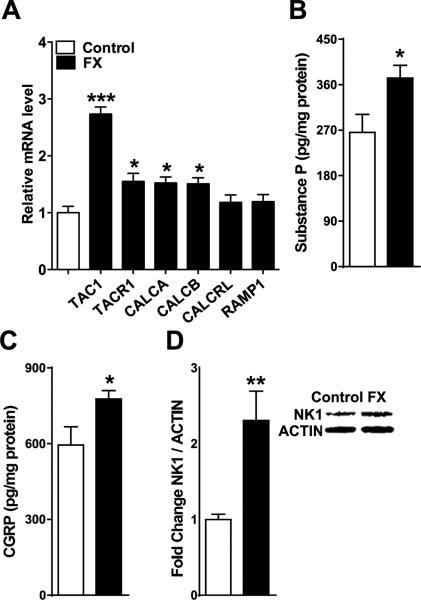
Figure 1. Distal tibia fracture facilitated spinal neuropeptide signaling in the ipsilateral L4,5 spinal cord segments at 4 weeks post-fracture (FX, n = 8), compared to control rats (Controls, n = 8).
(A) Tibia fracture caused increased spinal expression of the substance P (SP) gene (TAC1), the SP neurokinin1 (NK1) receptor gene (TACR1), the α-calcitonin gene-related peptide (α-CGRP) gene (CALCA), and the β-CGRP gene (CALCB). Fracture had no effect on the spinal expression of the CGRP calcitonin receptor-like receptor (CRLR) gene (CALCRL) or on the CGRP receptor activity-modifying protein (RAMP1) gene (RAMP1). Substance P (B) and CGRP (C) spinal protein levels, as measured by enzyme immunoassay, also increased at 4 weeks post-fracture. A western immunoblot assay was used to demonstrate a 2-fold increase in SP NK1 receptor protein levels after fracture (D). Values are means ± SE. One-way ANOVA (p < 0.001) with Bonferroni post hoc testing (A) and t-tests (B-D) *P< 0.05, ** P< 0.01 and *** P< 0.001 for FX vs Control.

