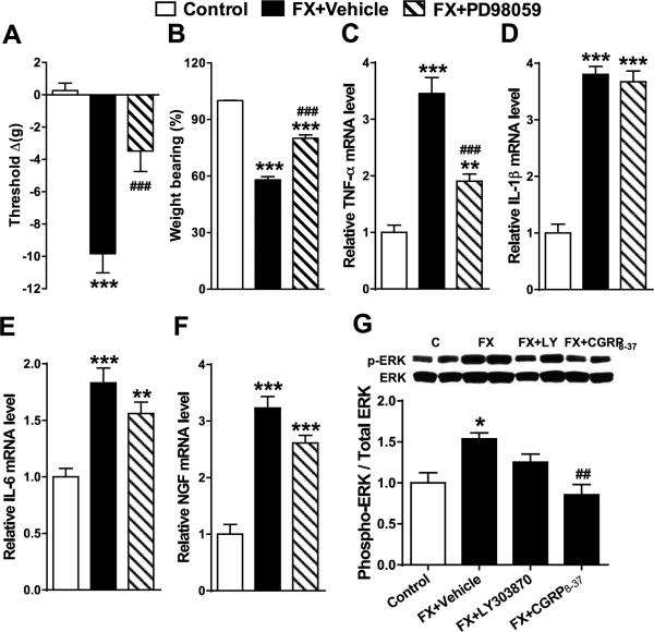
Figure 7. Spinal extracellular signal-regulated kinase 1/2 (ERK1/2) mitogen-activated protein kinase (MAPK) phosphorylation increased after fracture and contributed to upregulated spinal TNF-α expression and hindpaw nociceptive sensitization.
At 4 weeks post-fracture rats underwent intrathecal injection with vehicle (FX + Vehicle) or the ERK1/2 MAPK phosphorylation inhibitor PD98059 (20 μg). Peak analgesic effects for hindpaw von Frey allodynia (A) and unweighting (B) were observed at 1 hour post-injection. PD98059 intrathecal injection also partially inhibited post-fracture increases in spinal TNF-α mRNA levels (C), but had no effect on post-fracture increases in IL-1β (D), IL-6 (E) and NGF (F), compared to FX + vehicle group. At 4 weeks post-fracture there was an increase ratio of phosphorylated (pERK) to total ERK1/2 (ERK) in the lumbar spinal cord, indicating ERK1/2 activation (G). Intrathecal injection of the SP NK1 antagonist LY303870 (20μg) in fracture rats (FX + LY) did not significantly inhibit ERK1/2 phosphorylation, but injection of the CGRP antagonist CGRP8-37 (20μg) (FX + CGRP8-37) completely blocked post-fracture induced phosphorylation of ERK1/2. Values are means ± SE, n=8 per cohort. One-way ANOVA (p < 0.001) with Bonferroni post hoc testing *P< 0.05, ** P< 0.01 and *** P< 0.001 for FX + Vehicle, or FX + PD98059 vs Control, ### p< 0.001 for FX + PD98059, or FX + CGRP8-37 vs WT FX.

