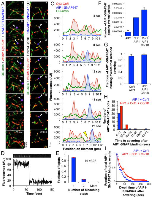
Figure 4. Recruitment of AIP1-SNAP647 to actin filaments induces rapid severing.
(A and B) Time points from TIRF movies (Supplementary Movie 5) showing Cy3-Cof1 and AIP1-SNAP647 binding to preassembled OG-labeled actin filaments after flow-in, both in the absence (A) and presence (B) of Cor1B. White arrowheads indicate appearance of AIP1-SNAP647 fluorescence; yellow arrowheads indicate severing. (C) Cy3-Cof1 and AIP1-SNAP647 spatiotemporal profiles; same filament as in (B). Green curve represents OG-actin fluorescence; yellow arrowhead indicates severing. (D) Stepwise photobleaching of surface-adsorbed AIP1-SNAP647. (E) Distribution of number of steps to complete photobleaching of AIP1-SNAP647 spots (± SE, shown in red, n=323). (F) Frequency of binding of AIP1-SNAP647 (± SEM, n=3) on actin filaments for the indicated conditions. (G) Fraction (± SEM, n=3) of AIP1-SNAP647 binding events that induce actin filament severing. No statistically significant difference was observed by chi-square test (p > 0.05). (H) Distribution of time intervals between appearance of AIP1-SNAP647 on filaments and severing. (I) Fitted curve for the distribution of AIP1-SNAP647 dwell times on actin filaments for the indicated conditions.

