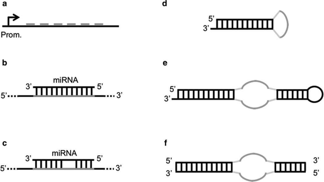
Fig. 2.
miRNA sponge constructs. (a) Basic sponge with six miRNA binding sites separated by 4-nt spacers. (b) Perfect miRNA binding site on a sponge. (c) Bulged miRNA binding site on a sponge. (d) Prototype decoy consisting of a short hairpin molecule where the loop exposes a binding site for an miRNA. (e) Tough decoy (TuD) with two exposed miRNA binding sites. (f) Synthetic tough decoy (S-TuD) consisting of two fully 2′-O-methylated RNA strands exposing an miRNA binding site each

