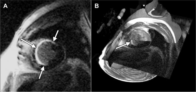
Figure 1.
A, Short-axis delayed contrast-enhanced MR image (5.4/1.3/175, 20° flip angle, 40-cm field of view, 256 × 160 matrix, 8-mm section thickness with 10-mm spacing) in 58-year-old man with extensive MI (arrows). B, Image in A overlapping 1H fast spin-echo MR image, with cut planes positioned to reveal the intersection of the two data sets on a line through the LV. Landmarks such as the epicardial and endocardial boundaries, aorta, apex, and base are used to guide manual registration of the partially transparent images, which are rendered stereoscopically. Arrow shows position of MI. Fast spin-echo image depicts signals from the three reference phantoms embedded in 23Na coil.

