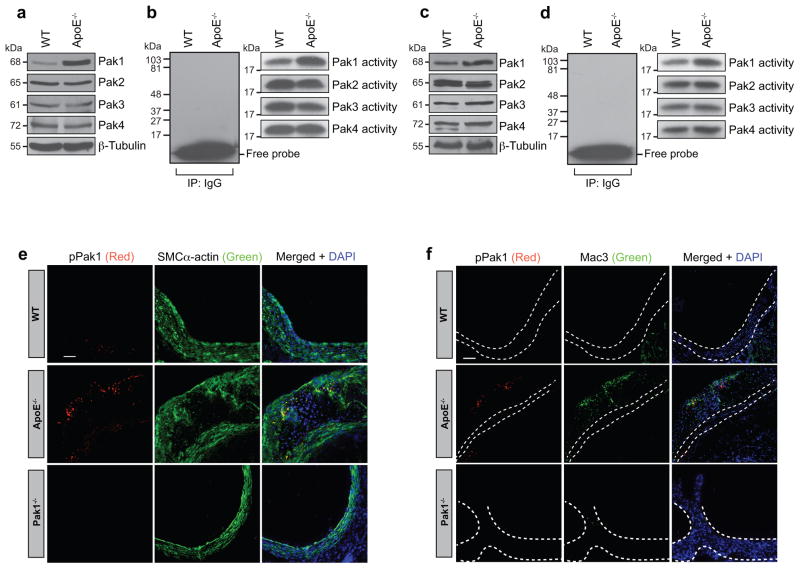
Figure 1. Pak1 expression and its activity increase in ApoE−/− mice.
(a–d) Aortic or primary peritoneal macrophage extracts of 24 week old C57BL/6 (WT) and ApoE−/− mice fed with chow diet were analyzed either by Western blotting for the indicated molecules using their specific antibodies (a and c) or an equal amount of protein from WT and ApoE−/− mice were immunoprecipated (IP) with rabbit IgG or anti-Pak1-4 antibodies and the immunocomplexes were assayed for kinase activity using myelin basic protein and [γ-32P]-ATP as substrates (b and d). (e, f) Aortic root cryosections of WT, ApoE−/− and Pak1−/− mice were stained by double immunofluorescence for pPak1 (red) and SMCα-actin or Mac3 (green). Scale bar in Figure 1e and f represent 50 μm.

