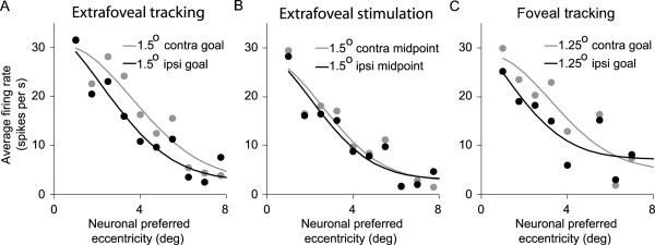
Figure 11.
Assessing shifts in the population of active neurons during extrafoveal tracking, extrafoveal stimulation, and foveal tracking with shifts in the retinotopic location of the midpoint between the two bars. (A) We performed an analysis similar to that of Figure 9 but for different retinotopic goal locations during extrafoveal tracking. We plotted the activity of our neurons when the goal was at a 1.5° eccentricity relative to gaze on either the contralateral (gray) or ipsilateral side (black) - along the axis of motion. Our neurons changed their activity with goal location (see Figures 2 and 5 for samples illustrating this) in a manner that was consistent with the entire center of mass of the active population shifting to reflect this goal location. (B) The same analysis on the same set of neurons revealed much smaller changes in neuronal activity during the extrafoveal stimulation condition when the monkeys could ignore the (invisible) midpoint. (C) During foveal tracking, we again observed shifts in the center of mass of the active population as the goal (now visible) shifted retinotopically. The smaller range of tracking errors in this condition meant that to have enough data to perform this analysis, we could only test for shifts associated with 1.25° goal locations as opposed to 1.5°.

