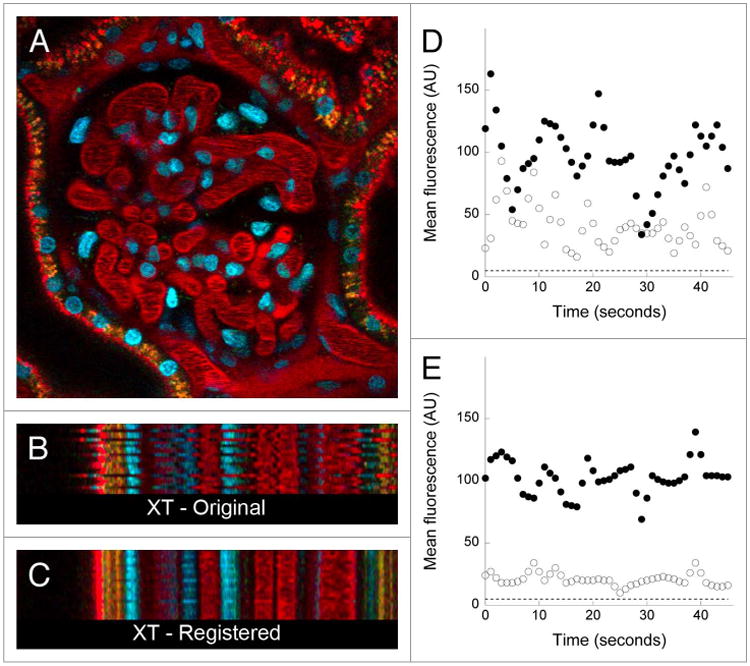
Figure 2.

Digital correction of motion artifacts in a time series of images collected from the kidney of a living rat. (A) First of 45 images collected from the kidney of a living rat, following intravenous injection of hoechst 33342 (labels nuclei blue) and texasred-labeled albumin. (B) Xt projection of the region identified by the horizontal line in Panel A, in which sequential linescans are arrayed vertically. (C) Xt projection of the same region shown in B, but after digital correction of the time series. image field is 160 microns across. the time series of the original and corrected image are presented in Video S2. (D and E) Quantification of mean fluorescence in a ten-pixel region located in the lumen of a glomerular capillary (closed circles), or in a region located 4 pixels (1.6 microns) away in the adjacent Bowman's space (open circles) before (D) or after (E), digital correction. dashed line—background signal level.
