
Fig. 3. N-α-PGP induces skin and pulmonary microvascular permeability.
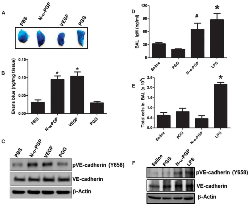
(A to C) Mice (n = 4 to 5 per group) were injected via the tail vein with Evans blue and then received abdominal subcutaneous injection of PBS alone, N-α-PGP (250 μg), PGG (250 μg), or VEGF (50 ng). Evans blue leak to the skin tissue was visually assessed (A) and quantified (B), and VE-cadherin phosphorylation after treatment was determined by Western blot (representative image) (C). *P < 0.05 versus PBS control by one-way ANOVA with Tukey post-test. (D to F) Mice (n = 4 to 5 per group) were intraperitoneally injected with saline alone or containing N-α-PGP (250 μg), PGG (250 μg), LPS (75 μg) once a day for 4 days, and then the total IgM in BAL fluid was measured by immunoassay (D) and total BAL cell (E), and VE-cadherin phosphorylation in lung lysate was measured by Western blot (F). *P < 0.05 versus saline or PGG, #P < 0.05 versus PGG by one-way ANOVA with Tukey’s multiple comparison post-test. All values represent means ± SEM.