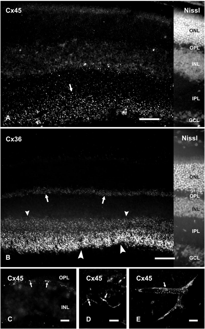
Figure 1.

Immunolabeling of Cx45 and Cx36 in adult mouse retina. A, B, Vertical retinal sections labeled by immunofluorescence, with the right portion of each field shown by counterstaining with fluorescent NeuroTrace. Punctate labeling with polyclonal anti-Cx45 is seen in the inner half of the IPL (A, arrow), and more sparsely in the outer half of the IPL (A). Labeling of Cx36 is very dense in the inner half of the IPL (B, large arrowheads), less dense in the outer half of the IPL (B, small arrowheads) and moderate in the OPL (B, arrows). C-E, Higher magnifications showing punctate labeling for Cx45 at the outer margin of the INL (C, arrows), and along blood vessels in the retina (D, arrows), and in cerebral cortex (E, arrow). ONL, outer nuclear layer; OPL, outer plexiform layer; INL, inner nuclear layer; IPL, inner plexiform layer; GCL, ganglion cell layer. Scale bars, 50 μm.
