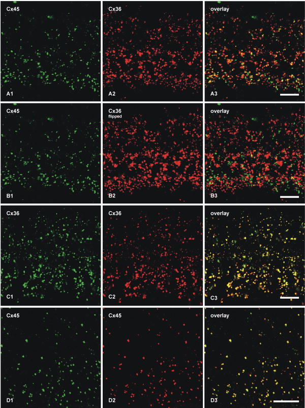
Figure 2.
Double-immunofluorescence images showing co-localization of Cx45 with Cx36 in vertical sections of the IPL of adult mouse retina. Images in A-D show the entire vertical span of IPL in single confocal scans. A, The same field (A1-A3) labeled with monoclonal anti-Cx45 and polyclonal anti-Cx36, showing Cx45-positive puncta labeled for Cx36 in retina after perfusion of mice with 20 ml of tissue fixative. B, The same set of confocal double immunofluorescence images as in A, showing minimal Cx45/Cx36 co-localization in the IPL after horizontal flipping of the image showing labeling for Cx36. C, The same field (C1-C3) of IPL double-labeled for Cx36 with monoclonal Ab37-4600 and polyclonal Ab36-4600, showing nearly total overlap of immunopositive puncta. D, The same field (D1-D3) in a tangential section through the IPL double-labeled for Cx45 with monoclonal MAB3101 and polyclonal Ab40-7000, showing nearly total overlap of immunopositive puncta. Scale bars, 10 μm.

