
Figure 1. Characterization of selected patient biopsies.
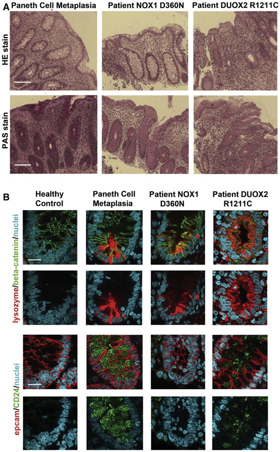
(A) HE and periodic acid–Schiff staining of colonic biopsy samples from an inflammatory bowel disease (IBD) control (Paneth cell metaplasia), a patient with the NOX1 D360N variant, and a patient with the DUOX2 R1211C variant. The patient with the NOX1 D360N variant shows focal inflammation, increased cellularity of inflammatory cells adjacent to normal area. The patient with the DUOX2 R1211C variant shows severe colitis with architectural distortion (crypt damage). Scale bar: 20 μm. (B) Immunofluorescence analysis with Paneth cell markers lysozyme and CD24 in colonic biopsy samples: lysozyme and EpCAM (red), β-catenin and CD24 (green), and nuclei (blue). Lysozyme was expressed in the crypts of the patients as well as the IBD control, but not in the healthy control. CD24 is expressed in colonic crypts in the IBD control and the patient with the DUOX R1211C variant, but neither in the healthy control nor the patient with the NOX1 variant. Scale bar: 10 μm.