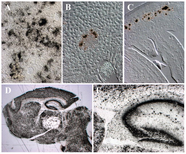
Figure 6. Aerosol infection of non-immunized and previously immunized mice with pathogenic RVFV.
Non-immunized mice show a severe hepatitis after aerosol exposure to ZH501 RVFV, while previously immunized mice show only mild hepatitis after aerosol exposure. (A–F) Differential interference contrast and in situ hybridization for RVFV RNA (black grains). (A) Non-immunized mouse shows severe hepatic infection at 6 days post-infection (DPI). Infection of the liver is delayed with aerosol infection compared to intraperitoneal infection. (B) Mice immunized with alphavirus replicons expressing the Gn glycoprotein of RVFV show occasional infected foci in the liver 3DPI. Virus is cleared from the liver by 6DPI. (C) 3DPI after aerosol exposure to RVFV, enteric infection is observed in epithelial cells at the depths of small bowel crypts in mice immunized with alphavirus replicons expressing the Gn glycoprotein of RVFV. (D& E) Despite modest systemic infection in mice previously immunized with DNA plasmids expressing Gn glycoprotein of RVFV fused to three copies of complement protein (C3d), aerosol exposure to RVFV ZH501 leads to lethal encephalitis 7 to 10 days later. (D) The brain of terminally ill mice illustrates that the vast majority of neurons are infected. (E) High power image of the hippocampus of (D).

