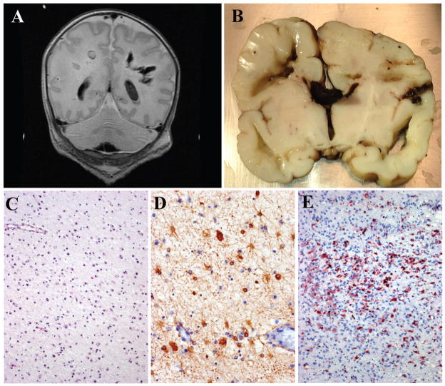
Figure 7. HPeV3 infection of neonates.
(A) T1-weighted non-contrast MRI from an HPeV3-infected infant. The child was healthy at birth but developed “neonatal sepsis” after exposure to an ill adult 30 days after delivery. Initial radiologic studies were normal; but after developing seizures, subsequent scans demonstrated cavitary deep white mater lesions. The infant died the following day. (B) Gross coronal section of the infant’s brain confirms the presence of deep-seated periventricular cavitary lesions with associated hemorrhage. (C) H&E stained sections adjacent to the cavitary lesions demonstrate a bland gliosis with mineralization and no adaptive immune cell infiltration. (D) Immunohistochemistry for GFAP confirms perilesional astrocytosis, while immunohistochemistry for CD68 confirms perilesional microglial activation (E).

