
Figure 4. Diverging and plane wave imaging of the displacements.
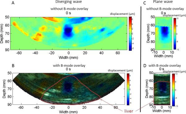
Peak negative HMI displacement imaging during HIFU ablation of a canine liver using diverging wave with no B-mode overlay (A), with B-mode overlay (B), using plane wave imaging with no B-mode overlay (C) and with B-mode overlay (D). The 50Hz HMI displacement sound corresponding to the ablation monitored with plane wave (Figure 4.C) was incorporated to the video.