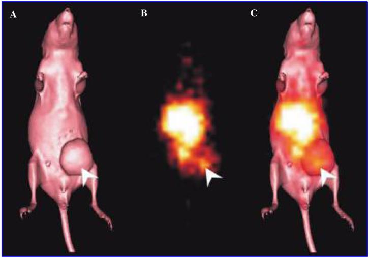
Figure 4.
(A) Surface-rendered computed tomography (CT) scan of a BALB/c nude mouse bearing a MCF-7 tumor on the left mammary line (right side of image). (B) Gamma-camera image obtained at 24 hours after an injection of the 111In-CHX-A″-DTPA hu3S193 multimer. The tracer uptake observed in the center of the image corresponds to blood pool and kidney uptake. Localization of the hu3S193 multimer to the MCF-7 tumor is indicated by a white arrow. (C) Overlayed gamma-camera image and CT surface render showing the localization of the multimer in the tumor (arrow).

