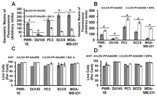
Figure 11. rLOX-PP-Atto565 uptake in bafilomycin A1 and EIPA treated cells.

Cells were pre-treated in the presence or absence of bafilomycin A1 (A) or EIPA (B), and rLOX-PP-Atto-565 uptake was measured by flow cytometry after 30 minutes of incubation. Bafilomycin decreased rLOX-PP-Atto565 uptake after 30 minutes. Data are means +/− SD, n = 3; *, p < 0,005. rLOX-PP uptake was inhibited by bafilomycin A, while it was stimulated by EIPA in all cell lines except possibly DU145 cells. In C and D, the LIVE/DEAD® Fixable Near-IR stain flow cytometry assay was used to determine the percentage of live cells. NT (dark gray bars), non-treated control cells; rLOX-PP-Atto565 (light gray bars), Bafilomycin A- or EIPA- + rLOX-PP-Atto565 (white bars).
