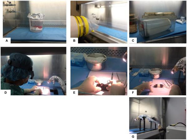
Figure 4. Skin transplantation procedure in GF mice.
A. Zone 1 with reusable cold pack submerged in a beaker of gluteraldehyde secured with rubber coated lead weight. B. Sterilzation cylinder with transport sleeve positioned to introduce GF mice and sterile caging material from flexible film isolator into Zone 1 of BSC. C. Sterile cage containing a sterile wide mouth Nalgene bottle introduced into Zone 1/Zone 2 used to transport post-operative mice from BSC to isolator. D. Surgeon wearing magnification visor preparing surgical site at left edge of Zone 2. E. Placement of skin graft on dorsal thorax of recipient GF mouse. F. Bandaged, anesthetized, post-operative recipient mouse. G. Fiber optic light positioned within the BSC in Zone 3 with sterile mylar film over light source attached to sterile orthopedic stockinette, covered with sterile aluminum foil and secured with sterile umbilical tape to sterile metal stand.

