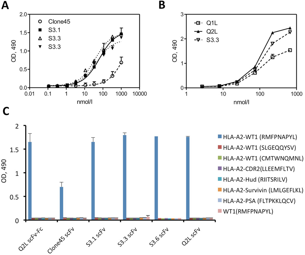
Figure 2. ELISA of scFv variants and Q2L scFv-Fc against HLA-A2-peptide monomers.
(a) Three scFvs (S3.1, S3.3 and S3.6) from the FACS sorting and parental Clone45 scFv were serially diluted and tested for binding to wells coated with HLA-A2-WT1 (RMFPNAPYL) monomer. (b) Q1L (single mutation), Q2L (double mutations) and S3.3 scFvs were serially diluted and added to wells coated with HLA-A2-WT1 (RMFPNAPYL) monomer. (c) The S3.1, S3.3, S3.6, Q2L, parental scFvs and Q2L scFv-Fc were serially diluted and added to wells coated with WT1 peptide (RMFPNAPYL), three types of HLA-A2-WT1 monomers and four irrelevant HLA-A2 monomers. Bound scFv or scFv-Fc were detected with an HRP-conjugated anti-Flag tag antibody or HRP-conjugated anti-human Fc antibody and the optical densities (O.D.) at 490 nm measured by Dynex MRX Microplate reader. For panels (a), (b) and (c), all samples were prepared in duplicate, and experiments were repeated 1–2 times with similar results. Values are shown as mean ± SD.

