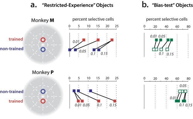
Figure 3.
IT selectivity among the “restricted-experience” objects (a) and “bias test” objects (b) for each monkey. a) Red points indicate the fraction of IT neurons selective among the “restricted-experience” objects at the trained position (counter-balanced across the two monkeys as shown). Blue points show selectivity among the same object set at the eccentricity-matched, non-trained position. Selectivity was determined by ANOVA (see Methods) and a range of significance levels (p values) used for deeming a neuron to be “selective” are indicated. Note that, in all cases, both monkeys showed a tendency for more selectivity at the trained position (red), relative to the non-trained position (blue). b) Same conventions as (a) for the “bias test” objects (Fig. 1a) presented at the same two retinal positions (filled green squares indicate the position that was trained with the “restricted experience” objects). Please note that, because the “bias test” objects were chosen to have greater within-object-set shape differences than the “restricted experience” object set (see Methods), it is unsafe to compare the absolute fraction of neurons showing shape selectivity among the two object sets.

