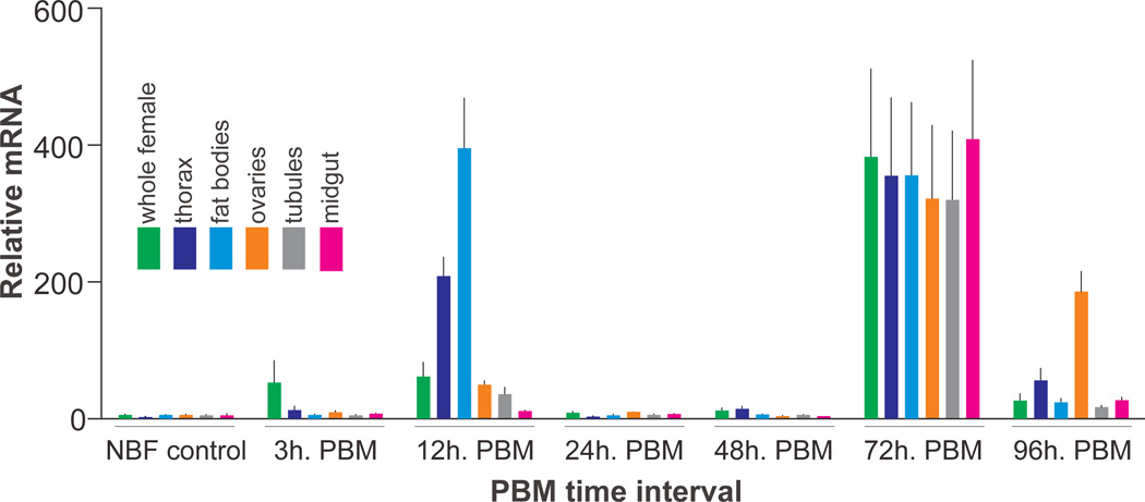
Fig. 5. AaSlif expression and regulation in selected mosquito tissues and organs.
The transcript levels were determined using quantitative PCR (qPCR). The data represent relative quantities of AaSlif transcript that were normalized with qPCR levels of ribosomal protein S7 (rpS7) mRNA in the same tissue sample set. Bars are means + SEM for n = 3 replicates collected from different sets of mosquitoes (ANOVA followed by Tukey’s HSD post hoc test). The RNA samples were isolated from whole body and various body parts and organs of adult female mosquitoes grouped as shown by the color-coding insert. The horizontal scale indicates group-specific conditions: non-blood fed (NBF, control), 3, 12, 24, 48, 72, and 96 h post-blood meal (PBM).

