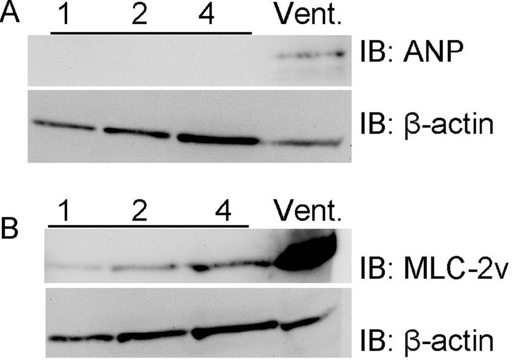
Figure 2. Aortic valve regions do not express ventricular myocardium markers.
Samples were prepared with increasing numbers of pooled embryonic mouse aortic valve regions, ranging from 1 to 4 valve regions per sample, or with a sample of the right ventricle. Samples were processed as described in Figure 1 and blotted for the ventricular myocardium markers ANP (A) and myosin light chain (MLC)-2v (B). ANP is completely restricted to the ventricular sample, and MLC-2v is expressed at extremely low levels compared with the ventricular sample. β-actin is shown as a loading control.

