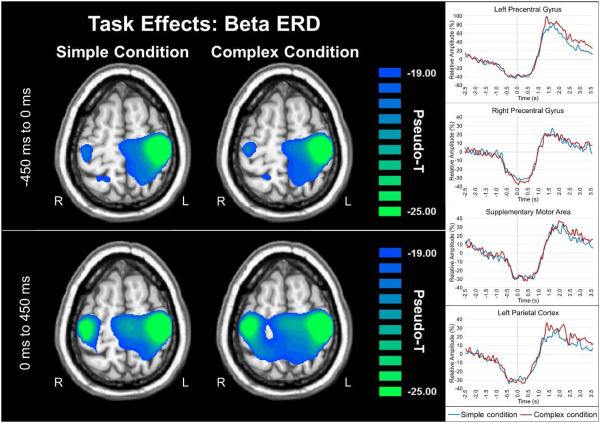
Figure 4. Beta ERD during movement planning and execution.
Group mean beamformer images (pseudo-t) of beta activity during movement planning (top left) and execution (bottom left) time bins are shown with virtual sensor data to the right. During both simple and complex movements participants exhibited significant beta ERD task effects in the bilateral primary motor cortex (stronger in the contralateral), superior parietal regions, cerebellum (not shown) and the SMA during both time windows (all p’s < .0001, corrected). To more precisely examine the dynamics, virtual sensors from the peak voxel in each region were extracted (right panel), which further confirmed the temporal progression of neuronal activity that was discernable in the discrete beamformer images. For each voxel time series, relative amplitude (in percentage from baseline) is shown on the y-axis, with time (in seconds) on the x-axis. The simple condition is shown with a blue line, while the complex condition is shown in red. Axial slices are shown in radiologic convention (right = left).

