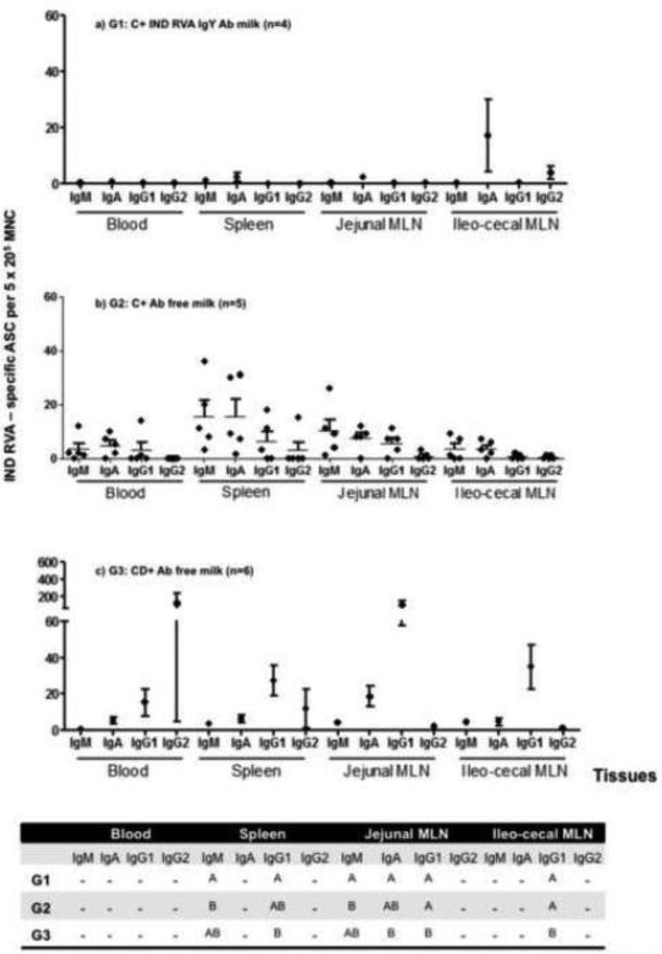
Figure 3.
Mean numbers of bovine RVA ASC per 5 × 10⁵/MNC obtained from systemic lymphoid tissues (PBL, spleen and MLN draining the small and large Intestine), at 21 PID. G1: calves fed with colostrum (C) followed by bovine RVA egg yolk supplemented milk with a final bovine RVA IgY Ab titer of 4096 for 7 consecutive days once diarrhea appearance; G2: calves receiving C followed by non-supplemented RVA Ab free milk; G3: colostrum deprived calves receiving RVA Ab free milk. All calves were orally inoculated with virulent Indiana RVA strain at 0 PID. For each tissue, when comparing mean ASC numbers of the same isotype, among treatment groups: different letter indicate a significant difference (Kruskal-Wallis rank sum test, p < 0.05). n = number of calves in each group. Error bars indicate SD. The “–” indicates no significant differences between groups.

