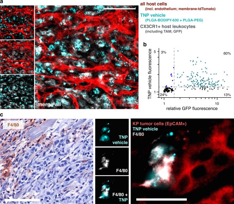
Figure 6. Tumor-associated CX3CR1+ and F4/80+ host phagocytes accumulate TNP vehicle in a syngeneic model of lung cancer.
Kras mutant p53−/− (KP) lung cancer cells were subcutaneously implanted into Cx3cr1GFP/+ reporter mice for directly visualizing GFP+ monocytes, dendritic cells, and TAMs. Tumor-adjacent stromal regions were imaged 24 hr following i.v. injection with TNP vehicle. Reporter mice expressed membrane-anchored tdTomato in all host cells, most visibly here with endothelium. Scale bar = 50μm. (b) Fluorescence intensities of GFP and TNP vehicle were quantified across single-cells based on a, showing most stromal cells that accumulate TNP vehicle were also GFP+ (n=4 animals, n=250 cells). (c) Histological analysis of F4/80+ host phagocytes, 24 hr post-injection with TNP vehicle. Immunohistochemistry (left) shows tumors stained with hematoxylin and F4/80 (brown ; scale bar = 100μm), and corresponds with immunofluorescence (right) showing F4/80+ host phagocytes, EpCAM+ KP tumor cells, and local accumulation of TNP vehicle (scale bar = 25 μm). F4/80 labels a subset of GFP+ cells in Cx3cr1GFP/+ reporter mice37 (namely, macrophages).

