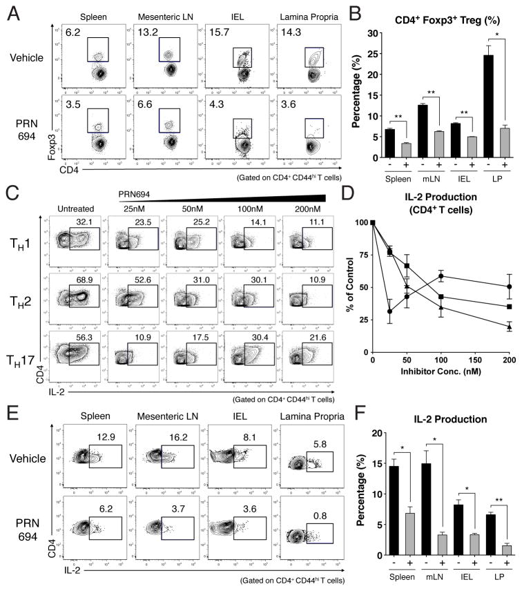
Figure 5. PRN694 treatment decreases Treg frequencies and IL-2 production from CD4+ T cells in vitro and in vivo.
(A–B) CD4+ T cells from spleen, mLN, IEL, and lamina propria from vehicle-treated or PRN694-treated mice at 7 weeks of post-transfer were examined for Foxp3 expression. (A) Dot plots show CD4 vs. Foxp3 staining, with numbers indicating the percentages of CD4+ Foxp3+ Treg cells, and (B) the graph shows a compilation of data from 3–5 mice in each group. (C–D) Naïve CD4+ T cells were stimulated in TH polarizing conditions with PRN694 at the indicated concentrations for 72hr. Cells were then re-stimulated with PMA and Ionomycin for 5hr and then analyzed for IL-2 production. (C) The percentages of IL-2-producing CD4+ T cells (gated on CD4+ CD44hi) are shown. (D) Compilation of data from 3 independent experiments is shown, with the data for each TH subset normalized to the percentage of positive cells in the absence of inhibitor. (E–F) Isolated lymphocytes from vehicle-treated or PRN694-treated mice at 7 weeks post-transfer were stimulated with PMA and Ionomycin, and analyzed for IL-2 production. Plots show gated CD4+ CD44hi T cells, and the graph shows a compilation of data from 3–5 mice in each group.

