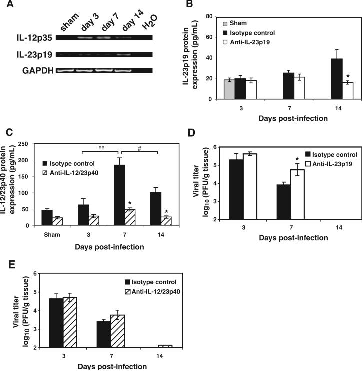
FIG. 3.
Antibody blocking of either IL-23p19 or IL-12/23p40 does not affect the ability to control viral replication within the brain. (A) RT-PCR revealed that intracranial instillation of MHV results in detection of transcripts specific for IL-23p19 and IL-12p35 in the brain at the indicated times post-infection. Each lane is from the brain of a single mouse and is representative of n 2−3 for each time point. ELISA indicating that both IL-23p19 (B) and IL-12/23p40 (C) are detected within the brains of MHV-infected mice at the defined time points post-infection (n 3−5 mice for each time point). Levels of IL-12/23p40 within the isotype control-treated mice were significantly elevated within the brain at day 7 post-infection when compared to levels present at day 3 (**p ≤ 0.007) and day 14 (#p ≤ 0.03) post-infection. Neutralizing antibody treatment significantly reduced (p ≤ 0.05) cytokine protein levels during peak expression in the brain compared to isotype control treatments at the indicated times post-infection. Treatment of MHV-infected mice with neutralizing antibodies specific for either IL-23p19 (D) or IL-12/23p40 (E) does not impair control of viral replication (PFU/g tissue) from the CNS. Data are presented as average SEM and represent a minimum of two independent experiments with a total n 5−9 for each antibody treatment group per time point examined. *p ≤ 0.05 compared to mice treated with control antibody.

