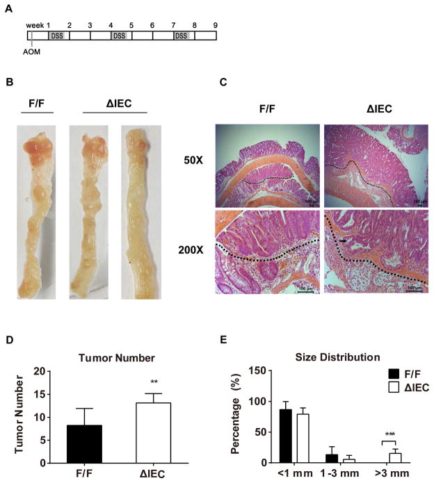
Figure 5. AOM plus DSS treatment causes an increased incidence and increased size of tumors in the colorectum of Ugt1ΔIEC mice compared to Ugt1F/F mice.
(A) A schematic schedule is shown as the AOM plus DSS feeding protocol. Each rectangle represents one week. (B) Photos of colon tumors in Ugt1F/F and Ugt1ΔIEC mice. (C) Representative tumor morphologies in colonic sections by H&E staining (50× and 200× magnification). The black arrow indicates the penetrating adenoma. (D) Tumor incidence and (E) size distribution in Ugt1F/F and Ugt1ΔIEC mice. At least seven mice were analyzed in each group, and results are presented as mean ± SD (* P<0.05, ** P<0.01, *** P<0.001).

