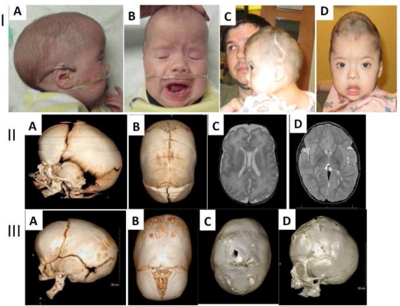
Fig.1. Case 1.

I: A, B; Photos of patient 1 before calvarial vault reconstruction. A. Photo shows typical compensatory scaphocephaly and micrognathia, as well as dysmorphic features including low set ears, frontal bossing of the cranial vault secondary to sagittal suture fusion. C,D; 24 months of age demonstrating turribrachycephaly. II: A, B; CT scan at birth showing the micrognathia (A) and the partial sagittal craniosynostosis (B). C, D; Axial T2 FLAIR MRI at birth shows adequate brain growth. Scan is notable for mild lissencephaly. III: A, B; CT scan at 5 months post mandibular distraction showing persistent micrognathia and progression of the sagittal craniosynostosis. C, D; CT scan at 24 months of age demonstrating craniocerebral mismatch with effacement of the sagittal suture and the coronal sutures.
