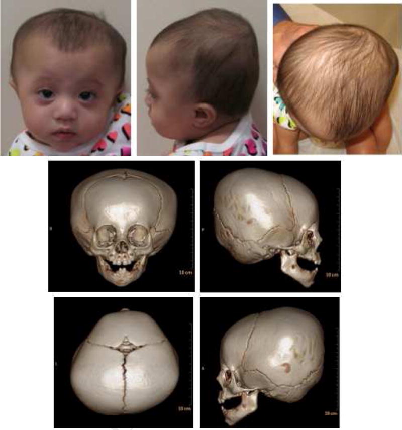
Fig.3. Case 3.

Three-dimensional reformatted CT scan imaging demonstrates head shape and lack of findings consistent with craniosynostosis and consistent with physiologic closure of the metopic suture. A. Frontal view; B. Right lateral view; C. Posterior view; D. Left lateral view. Note occipital flattening and biparietal peaking, physiologic closure of metopic suture and patency of other cranial sutures.
