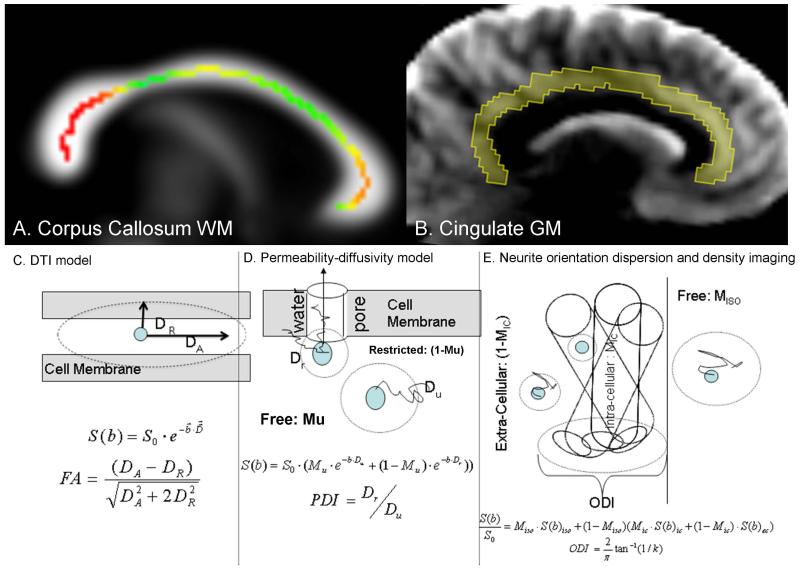
Figure 1.
Upper panel: (A) Corpus callosum (CC) white matter (WM) region-of-interest was identified by thresholding the FA image at FA=0.20. The skeleton of the midsagittal colossal WM measured using the ENIGMA-DTI pipeline is shown as colored line with the magnitude of FA values represented by colors and overlaid on the fractional anisotropy map at midline. (B) The region of interest for cingulate gray matter (GM) was identified using radial diffusivity maps that show excellent contrast between GM, WM and CSF. Lower panel: schematic comparison of the DTI model (C), permeability-diffusivity (PD) model (D) and Neurite orientation dispersion and density imaging (NODDI) model (E). The DTI model assumes that the signal is produced by a single pool of anisotropically diffusing water and characterizes this anisotropy using fractional anisotropy (FA). The PD-model, developed by Sukstanskii et al., (Sukstanskii, et al. 2004), assumes that the signal is produced by two quasi-pools of isotropically diffusing water. Unrestricted pool (Mu) is produced by water molecules that are sufficiently away from the cellular membranes to be unaffected by them. The water near the membrane forms the restricted compartment (1-Mu) whose diffusivity depends on both the passive diffusivity of water through the cellular/myelin membrane and the active (thick arrow) permeability via the ionic channels and water pores that use water as a substrate for compartment exchange. The NODDI model, developed by Zhang et al., (Zhang, et al. 2012) assumes contribution from three compartments: the unrestricted diffusion compartment (MISO), intra-cellular (MIC) and extra cellular (1- MIC). Neurites (neurons or axons) are expected to have a given angular distribution about the principle axis that is characterized by the orientation dispersion index (ODI).

