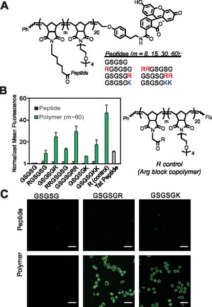
Figure 1.

Cellular internalization of GSGSG polymers and analogues. A) Chemical structure of peptide block copolymers. B) Flow cytometry data showing fluorescent signatures of HeLa cells treated with the polymers (m ~ 60) and their monomeric counterparts. All data are normalized to the vehicle (DPBS), which is assigned a value of 1. The R control is a block copolymer that contains a single Arg attached via a short linker at each polymer side chain of the first block (m ~ 60). “Flu” is the fluorescein end-label shown in A. C) Live-cell confocal microscopy images showing the average intensities from six consecutive 1 μm slices of HeLa cells treated with peptides and polymers (m ~ 60). Scale bars are 50 μm. In each study, the concentration of material is 2.5 μM with respect to fluorophore.
