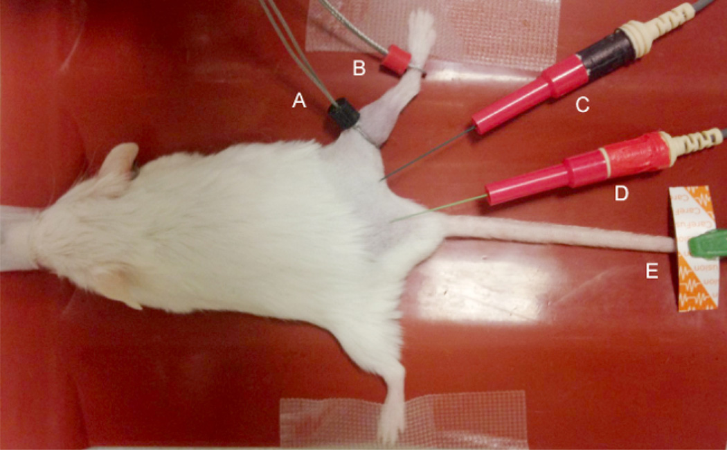
Figure 1. Electrode Placement.
The black (E1) “active” electrode (A) and red (E2) “reference” recording electrode (B) are placed over the gastrocnemius at the proximal portion of the gastrocnemius at the knee. The stimulating cathode (black) (C) and anode (red) (D) are inserted subcutaneously proximal to the recording electrodes to generate distal responses. A disposable disk electrode (D) is placed on the hind limb, tail or sacrum as a ground to minimize artifact.

