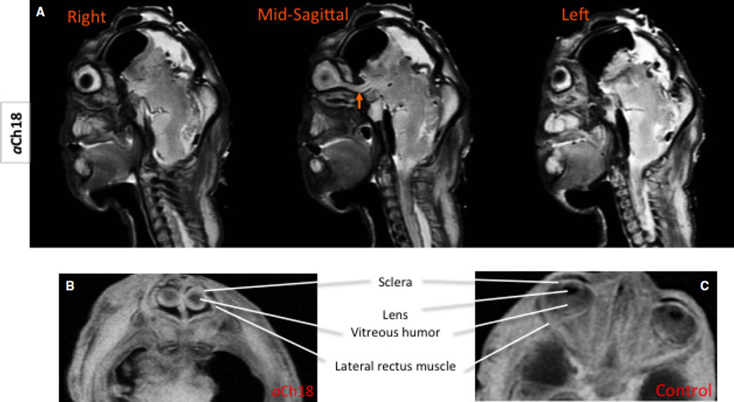
Fig. 4.
Eye structures in the aCh18 synophthalmic cyclops analysed with MRI. (A) Sagittal slices of the aCh18 fetus from right to left through the midline showing a fused optic nerve midsagitally (arrow); T2 weighted (B) horizontal slice through the mid-ocular region demonstrating the absence of the lens, and the presence of some extraocular muscles. (C) The control for (B) shows the normally developed intrinsic ocular structures at 29 weeks. (B and C) T1-weighted MRIs. Arrow demarcates medially located single optic nerve.

