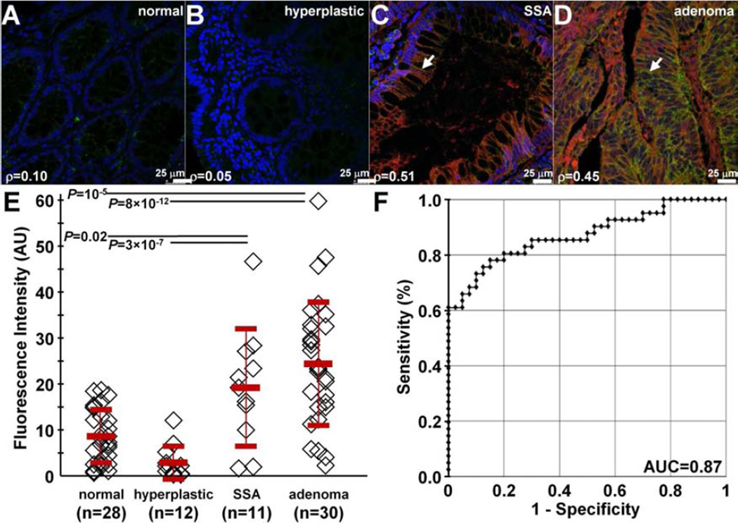
Fig. 9. Binding of claudin-1 peptide to human proximal colonic neoplasia.
On confocal microscopy, there was minimal binding of RTS*-Cy5.5 peptide (red) and AF488-labeled anti-claudin-1 antibody (green) to human A) normal colonic mucosa and B) hyperplastic polyps. DAPI (blue) identifies nuclei. Strong staining with both peptide and antibody was observed for representative specimens of C) sessile serrated adenomas (SSA) and D) adenomas from the proximal colon. The extent of co-localization of peptide and antibody binding is characterized by the Pearson’s correlation coefficient ρ. Representative images were selected from n = 28 normal, n = 12 hyperplastic polyps, n = 11 SSA, and n = 30 adenomas. E) We found a significantly greater mean (±std) intensity for adenomas (25.5±14.0) versus normal (9.1±6.0) and hyperplastic polyps (3.1±3.7), P=10−5 and 8×10⁻¹², respectively, as well as for SSA (20.1±13.3) versus normal and hyperplastic polyps, P=0.02 and 3×10⁻⁷, respectively. Analysis used an ANOVA models with means for 4 groups, fit to log-transformed data. The fluorescence intensities from 3 boxes (20×20 µm2) located randomly on cells within each specimen were measured and averaged. F) ROC curve shows an area under the curve of AUC = 0.87.

