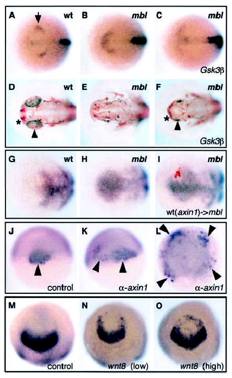
Figure 4.

Mbl/Axin1 functions both in anterior neural plate patterning and axis development. (A–F) Dorsal views of neural plates and brains of wild-type (A,D), mbl−/− (B,E), and mbl−/− embryos injected with gsk3 β RNA (C,F) stained for the expression of flh in the epiphysial region of the diencephalon (arrow, A) at bud stage (A–C) or stained with an α-acetylated tubulin antibody at pharyngula stage (D–F). The embryos injected with gsk3β RNA show partial rescue of thembl−/− phenotype as determined by the pattern of flh expression at bud stage and the presence of telencephalon (asterisk) and small eyes (arrowhead) at pharyngula stage. (G–H) Dorsal views of neural plates of wild-type (G), mbl−/− (H), and mbl−/− embryos in which wild-type cells expressing axin1 RNA were transplanted into the anterior neural plate at 70% epiboly stage (I) showing expression of fkd3 (characteristic of mid/caudal diencephalon identity in wild-type) at bud stage. Ectopic rostral fkd3 expression is absent in the transplanted cells and in some of the mbl−/− host cells adjacent to the transplanted cells. (J–L) Dorsal (J,K) and animal pole (L) views of a wild-type embryo and embryos injected with α-axin1 morpholino antisense oligonucleotides (K,L) stained for the expression of flh (a marker of organizer tissue, arrowheads) at shield stage. (J) An uninjected wild-type control embryo showing a single expression domain of flh within the dorsal organizer region. In (K), the injected embryo shows expansion of flh expression on the dorsal side of the embryo. In (L), the injected embryo has multiple sites of flh expression indicative of widespread dorsalization. (M–O) Animal pole views of bud-stage control embryo (M) or embryos injected with increasing (left to right) doses of wnt8 RNA (N,O). In addition to variably causing dorsalization (data not shown), pax2.1/noi expression (normally restricted to the presumptive midbrain), spreads into the anteriormost regions of the neural plate of injected embryos. This expansion is unlikely to be simply a result of dorsalization of the ectoderm, as inhibition of BMP signaling also dorsalizes ectoderm but does not lead to anterior expansion of pax2.1 expression (e.g., Barth et al. 1999).
