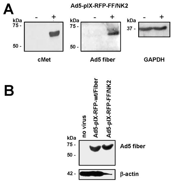
Figure 2.
Western blot analysis of NK2 expression in HEK293 cells infected with Ad5-pIX-RFP-FF/NK2. Notes: (A) Western blot analysis of viral HGF/NK2 and Ad5 fiber expression in uninfected HEK293 cells and HEK293 cells infected with Ad5-pIX-RFP-FF/NK2; analysis of cellular GAPDH expression was performed as a loading control. (B) Western blot analysis of viral Ad5 fiber expression in uninfected HEK293 cells and HEK293 cells infected with either Ad5-pIX-RFP-wt/Fiber or with Ad5-pIX-RFP-FF/NK2; analysis of cellular β-actin expression was performed as a loading control. The cells were infected with Ad5-pIX-RFP-wt/Fiber or Ad5-pIX-RFP-FF/NK2 at an MOI of 50 VP/cell. Untreated cells were included as a control. At 2 days following treatment, protein lysates were isolated and boiled for 5 minutes at 95ºC. Equal protein lysates were separated on 10% SDS-PAGE gels. The proteins were then transferred onto PVDF membrane, blocked, and probed with specific monoclonal antibodies for HGF-α or Ad5 fiber. An anti-GAPDH or anti-β-actin antibody was used as loading controls. Abbreviations: MOI, multiplicity of infection; VP, viral particles; SDS-PAGE, sodium dodecyl sulfate-polyacrylamide gel electrophoresis; PVDF, polyvinylidene fluoride; HGF, hepatocyte growth factor; GADPH, glyceraldehyde 3-phosphate dehydrogenase; NK2, a secreted truncated splicing variant that extends through the second kringle domain; HEK293, human embryonic kidney cell line; Ad5, adenovirus serotype 5.

