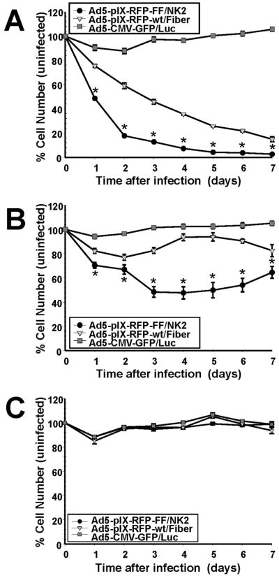
Figure 8.

Ad5-pIX-RFP-FF/NK2 oncolysis assay. Notes: Each cell line, including Hep3B2.1 (A), SNU-449 (B), and MCF-7 (C), was seeded in replicates of six into 96-well plates using 3×10³ cells/well. The next day, the cells were infected with Ad5-pIX-RFP-FF/NK2, Ad5-pIX-RFP-wt/Fiber, or a non-replicating Ad5-CMV-GFP/Luc virus, at an MOI of 1,000 VP/cell, and incubated at 37ºC for 7 days. At each time point, cell survival was determined in replicate wells using a CellTiter-Blue cell viability assay. Each experimental condition was performed in triplicate, and the experiments were repeated three times. The results were expressed as the mean of the three experiments ± SEM. Statistical significance was determined using a two-tailed Student’s t-test; differences were considered statistically significant (*) if P<0.05. Abbreviations: MOI, multiplicity of infection; VP, viral particles; SEM, standard error of the mean; Ad5, adenovirus serotype 5; NK2, a secreted truncated splicing variant that extends through the second kringle domain.
