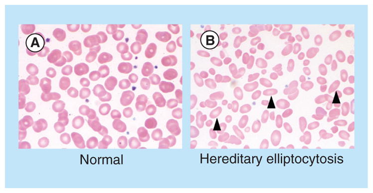
Figure 3. Morphologic features of normal red blood cells and congenital red blood cell defects.

Images demonstrate the peripheral blood smear from a (A) normal patient, and a (B) patient with hereditary elliptocytosis. Arrowheads highlight cells with characteristic morphologic defects.
