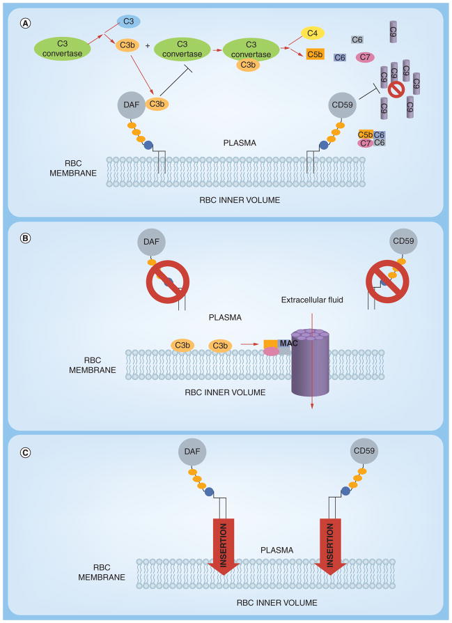
Figure 5. Approaches to ‘painting’ of red blood cells with protective complement regulatory proteins.
(A) Normal RBCs expresses complement-regulatory proteins including decay accelerating factor (DAF) and CD59 which are tethered to the RBC lipid bilayer of the cell membrane by a Glycosylphosphatidylinositol (GPI) anchor.
The presence of both DAF and CD59 protects the cells from homologous complement-mediated lysis. (B) Paroxysmal nocturnal hemoglobinuria RBCs are more vulnerable to complement mediated lysis due to a reduction or complete absence of GPI-anchored DAF and CD59. Continuous complement activation leads to the formation of membrane attacking complex which forms pores in the RBC membrane, allowing extracellular fluid to the enter, resulting in the swelling and ultimately rupture of the RBC. (C) RBC ‘painting’ is a method of incorporating proteins such as DAF and CD59 via GPI anchors into the membrane, decreasing the RBC sensitivity towards complement-mediated lysis.

