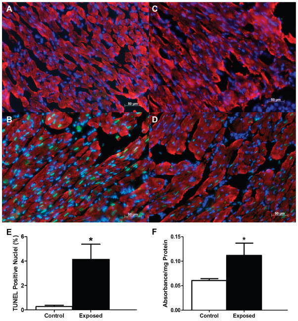
Fig. 1.
Cardiac apoptotic index following mountaintop removal mining particulate matter (PMMTM) exposure. Representative fluorescent images of cardiac tissue. A: negative control with exclusion of TdT enzyme. B: positive control with inclusion of DNase I. C: control with vehicle instillation. D: exposure with PMMTM instillation. DAPI-stained nuclei are indicated in blue, whereas terminal dUTP nick-end labeling (TUNEL)-positive nuclei are shown in green, and red indicates heavy chain cardiac myosin. Scale bar = 50 μm. E: apoptotic index was calculated as the percentage of total cardiomyocyte nuclei that were TUNEL-positive nuclei. Values are means ± SE; n = 3 for each group. F: relative cytosolic histone concentrations determined by ELISA on left ventricular tissue from control and PMMTM-exposed animals. Values are means ± SE; n = 5 for each group. *P < 0.05 for control vs. exposed.

