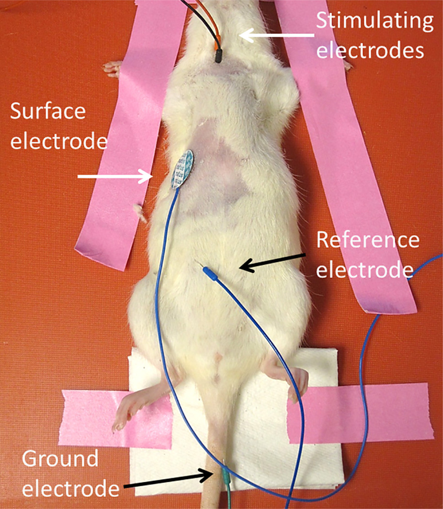
Figure 1. CMAP electrode placement.
The ground electrode is placed subcutaneously into the tail. The reference electrode is placed into the abdomen. The surface recording electrode is placed transversely along the unilateral costal margin. Stimulating electrodes are depicted as both red and black. Please click here to view a larger version of this figure.

