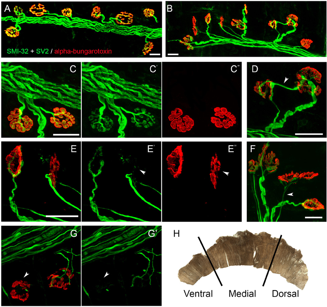
Figure 4. NMJ phenotypes.
Confocal imaging was conducted to visualize post-synaptic acetylcholine receptors (in red labeled with rhodamine-conjugated α bungarotoxin) and axonal fibers and pre-synaptic terminals (in green labeled with SMI-32 and SV2 antibodies, respectively). All NMJs in hemi-diaphragm were completely intact in control non-diseased wild-type rats (A, C). On the contrary, SOD1G93A rats (a rodent ALS model) showed significant pathology at hemi-diaphragm NMJs (B), including complete denervation (G, arrowhead), partial denervation (E, arrowhead; G, NMJ on right), multiple innervation (D) and thin pre-terminal axons (F, arrowhead). The diaphragm muscle is sub-divided into three separate sections for NMJ morphology analysis (H)13. Scale bar: 25 µm. Please click here to view a larger version of this figure.

