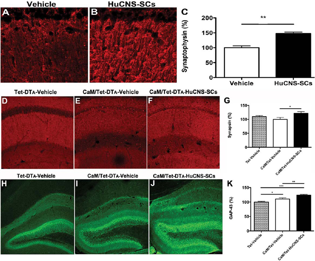
FIGURE 6.
HuCNS-SC transplantation increases presynaptic markers and axonal sprouting. (A) Immunolabeling of synaptophysin in vehicle injected 3xTg-AD mice. (B) HuCNS-SC injection results in a 47% increase in synaptophysin levels in the stratum radiatum of CA1, quantified in (C, N = 5, t-test P < 0.008). (D) Synapsin in control unlesioned mice reveals a typical pattern of presynaptic innervation within the stratum radiatum of CA1. (E) Surprisingly, synapsin levels are only slightly diminished in lesioned mice. (F) In contrast, HuCNS-SC transplantation significantly increases the density of presynaptic innervation, quantified in (G, N = 8–12, ANOVA P < 0.05, Fishers PLSD P = 0.016). (H) Compared to control unlesioned mice, we also detected a significant increase in GAP-43 immunoreactivity within the dentate gyrus of lesioned CaM/Tet-DTA mice with vehicle injections (I). More importantly, we observed a further enhancement of GAP-43 expression in lesioned mice transplanted with HuCNS-SC (J), quantified in (K). (N = 10, ANOVA P < 0.05, Fishers PLSD P = 0.028, P = 0.006, P = 0.0001). Data presented as mean- ± SEM. [Color figure can be viewed in the online issue, which is available at wileyonlinelibrary.com.]

