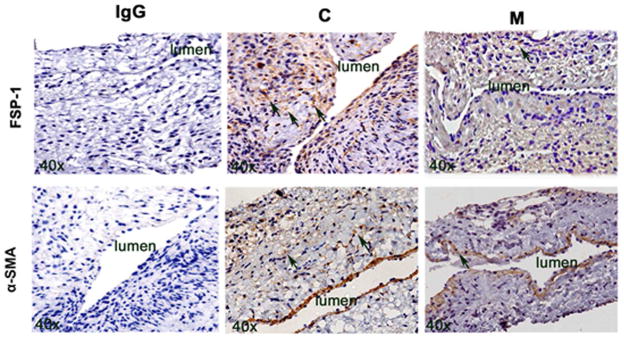
Figure 5.
FSP-1 and α-SMA staining in murine AVF at day 7 and 21 after placement of AVF in outflow vein alone (C) and MSC transplanted vessels (M). Upper panel is the representative sections after FSP-1 staining in the venous stenosis of the M transplanted and C vessels at day 21. Brown staining cells are positive for FSP-1 (arrow). IgG negative controls are shown. All are original magnification X 40. Lower panel is the representative sections after α-SMA staining in the venous stenosis of the M transplanted and C vessels at day 21. Brown staining cells are positive for α-SMA (arrow).

