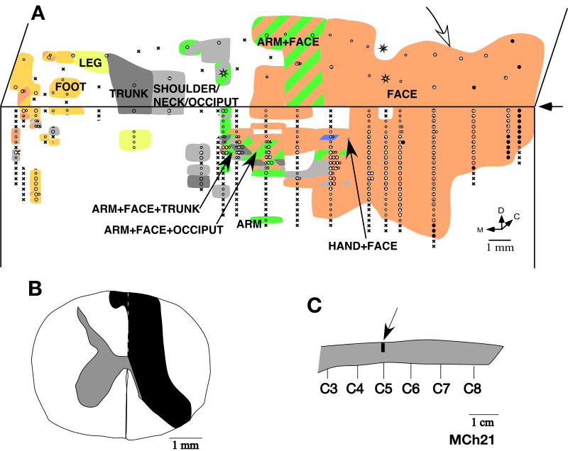
Fig. 12.
(A) Somatotopy in the left area 3b and the bordering cortex in monkey MCh21. The white arrow on the top marks the expected location of the normal hand-face border as estimated from the location of the tip of the intraparietal sulcus (see Fig. 15). Note the expansion of the face responsive regions into the deafferented hand, arm and occiput areas. Receptive fields on the face were also seen in the medial most region intermingled with the leg and the foot representations. Conventions as for Fig. 4A. (B) Reconstruction of the spinal cord lesion on a coronal section (region shown in black). (C) Location of the lesion (arrow) on a dorsal view of the spinal cord. Rostral is to the right.

